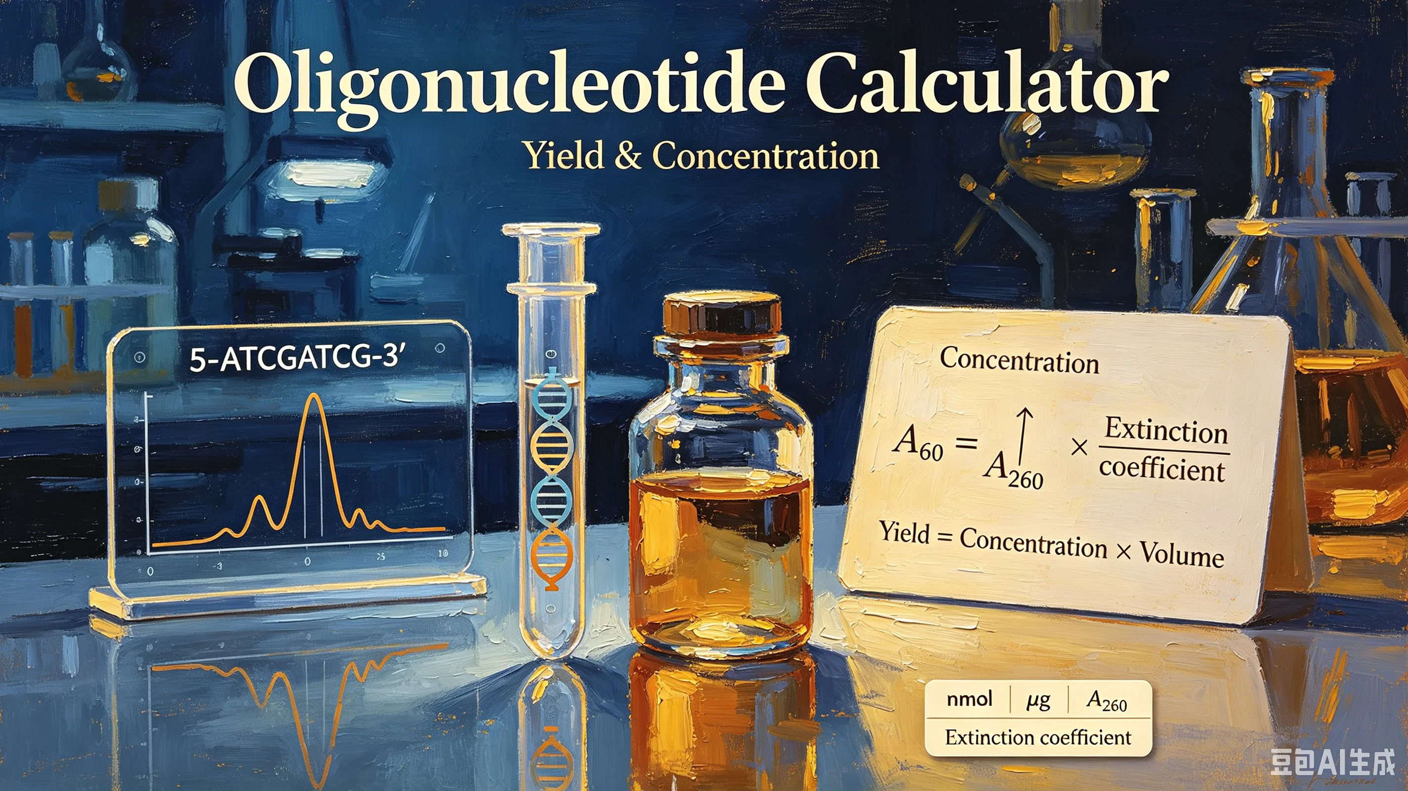
Calculate oligo concentration from OD260, yield in nmol and μg, molecular weight, and resuspension volume. Covers extinction coefficients, the 33 μg/mL rule, nearest-neighbor vs. base-sum methods, and primer dilution for PCR. Free oligo calculator included.
You just received a lyophilized primer from the synthesis company. The tube says "4.2 OD, 21.3 nmol" — but what does that mean, and how much buffer do you add to make a 100 μM stock? Oligonucleotide calculations trip up even experienced researchers because they involve three linked quantities: concentration (from UV absorbance), yield (total amount synthesized), and molecular weight (sequence-dependent). Get one wrong and your PCR fails, your qPCR standard curve is off, or your experiment wastes expensive oligos. This guide explains the math, the shortcuts, and a step-by-step protocol for calculating everything from an OD reading.
The Three Quantities You Need
Every oligo calculation revolves around three numbers:
1. Concentration (C) — how much oligo is in a given volume, usually in μM (micromolar) or μg/mL. Measured by UV absorbance at 260 nm.
2. Yield (amount) — total oligo in the tube, usually in nmol (nanomoles) or μg (micrograms). This is what the synthesis company reports. Yield = Concentration × Volume.
3. Molecular weight (MW) — the mass of one mole of your specific oligo sequence, in g/mol. Depends on length and base composition. Needed to convert between molar (nmol, μM) and mass (μg, μg/mL) units.
The challenge: UV spectrophotometers give you OD260 (absorbance), not concentration directly. To get concentration, you need the extinction coefficient — and that depends on your oligo's sequence.
Step 1: Calculate Concentration from OD260
DNA and RNA absorb UV light maximally at 260 nm. The relationship follows the Beer-Lambert law:
A₂₆₀ = ε × C × l
Where A₂₆₀ = absorbance at 260 nm (OD260), ε = extinction coefficient (M⁻¹cm⁻¹), C = concentration (M), and l = path length (cm, usually 1 cm).
Rearranged:
C (M) = A₂₆₀ / ε
C (μM) = A₂₆₀ × 10⁶ / ε
The extinction coefficient (ε) depends on the bases in your oligo because each base absorbs 260 nm light differently:
Base | Extinction Coefficient (M⁻¹cm⁻¹) |
|---|---|
dA (deoxyadenosine) | 15,400 |
dG (deoxyguanosine) | 11,500 |
dC (deoxycytidine) | 7,400 |
dT (deoxythymidine) | 8,700 |
Simple method (base sum): Add up the extinction coefficients of each base in your sequence. For a 20-mer with 5 A, 5 G, 5 C, 5 T:
ε = (5×15,400) + (5×11,500) + (5×7,400) + (5×8,700) = 215,000 M⁻¹cm⁻¹
Accurate method (nearest-neighbor): Bases stacked adjacent to each other have slightly different absorbance due to base-stacking interactions. The nearest-neighbor model accounts for this and is ~2–5% more accurate. Most online calculators use this method. For routine lab work, the base-sum method is usually sufficient.
The "33 rule" shortcut: For single-stranded DNA oligos, a widely used approximation is:
C (μg/mL) = OD₂₆₀ × 33
This works because the average μg/mL per OD260 for a random ssDNA oligo is ~33. It's quick but ignores sequence composition — an A-rich oligo (high ε) will have a different factor than a C-rich oligo (low ε). For precise work, always calculate ε from the sequence.
RNA oligos: Use different extinction coefficients (A: 15,000, G: 12,000, C: 7,000, U: 9,900) and the "40 rule" (C = OD260 × 40 μg/mL) as a rough approximation.
Step 2: Calculate Molecular Weight
To convert between molar units (μM, nmol) and mass units (μg/mL, μg), you need the molecular weight of your specific oligo.
Method: Sum of base weights minus water.
When nucleotides are linked by phosphodiester bonds, each bond releases one water molecule (H₂O = 18.02 g/mol). So:
MW = Σ(base MW) − (n−1) × 18.02 + terminal groups
Where n = number of bases, and terminal groups depend on modification:
Standard oligo with 5'-OH and 3'-OH: add ~18.02 (one water for the free ends)
5'-phosphorylated: add ~79.98 (phosphate group)
Common modifications (FAM, biotin, etc.) add their own MW
Average base molecular weights for DNA:
Base | MW (g/mol) |
|---|---|
dA | 313.21 |
dC | 289.18 |
dG | 329.21 |
dT | 304.20 |
Quick approximation: For a random DNA oligo, average MW per base ≈ 309 g/mol. So:
MW ≈ (n × 309) + 79 (for 5'-phosphate) or + 18 (for 5'-OH)
Example: 20-mer, 5'-OH: MW ≈ (20 × 309) + 18 = 6,198 g/mol ≈ 6.2 kDa.
For precise work, calculate from the actual sequence. A 20-mer that's all A has MW ~6,282; all C is ~5,802 — a 8% difference.
Step 3: Calculate Yield
Once you have concentration and volume, yield is straightforward:
Yield (nmol) = C (μM) × V (mL)
Yield (μg) = C (μg/mL) × V (mL)
Or, if you know molar yield and MW:
Yield (μg) = Yield (nmol) × MW (g/mol) / 1000
Example: Your oligo (MW = 6,200 g/mol) is resuspended in 500 μL (0.5 mL). You measure OD260 = 0.42. ε = 200,000 M⁻¹cm⁻¹.
C (μM) = 0.42 × 10⁶ / 200,000 = 2.1 μM
Yield (nmol) = 2.1 × 0.5 = 1.05 nmol
Yield (μg) = 1.05 × 6,200 / 1000 = 6.51 μg
The synthesis company typically reports yield in both nmol and OD units. 1 OD of a typical 20-mer ≈ 5 nmol ≈ 33 μg (using the 33 rule).
Step 4: Resuspension & Dilution
This is the calculation people do most often: "I have X nmol of oligo. How much buffer do I add to make a Y μM stock?"
V (μL) = Yield (nmol) / Target C (μM) × 1000
Example 1: Make a 100 μM stock from 21.3 nmol
V = 21.3 / 100 × 1000 = 213 μL
Add 213 μL of buffer (TE or nuclease-free water), vortex, and you have a 100 μM stock.
Example 2: Make a 10 μM working solution from 100 μM stock
Use C1V1 = C2V2:
V₁ = (C₂ × V₂) / C₁ = (10 × 1000) / 100 = 100 μL
Add 100 μL of 100 μM stock to 900 μL buffer = 1 mL of 10 μM working solution.
Typical primer stocks: Most labs store primers at 100 μM (100 pmol/μL) and use them at 0.2–1 μM final in PCR. A 20 nmol yield gives 200 μL of 100 μM stock — enough for ~200–1000 PCR reactions depending on volume.
Step-by-Step Lab Protocol
Step 1: Find your oligo's specs.
Locate the synthesis report. It should list: sequence, length, OD260, yield (nmol and/or μg), and sometimes MW and extinction coefficient.
Step 2: Calculate MW and ε (if not provided).
Use the base-sum method for ε and the base-weight method for MW. Or use an online calculator.
Step 3: Resuspend the oligo.
Add the calculated volume of buffer (usually TE pH 8.0 or nuclease-free water) to the lyophilized pellet. Vortex 10–15 seconds, spin down briefly.
Step 4: Verify concentration by OD260.
Dilute a small aliquot (e.g., 2 μL stock + 98 μL water = 1:50 dilution) and measure A260. Calculate:
Stock C (μM) = (A₂₆₀ × dilution factor × 10⁶) / ε
Step 5: Compare with expected.
If measured concentration is within 10–15% of expected, you're good. If it's way off, you may have pipetting error, incomplete resuspension, or the company's yield estimate was off.
Step 6: Make working dilutions.
Dilute the 100 μM stock to 10 μM working solution (or directly to your assay concentration). Aliquot and store at -20°C to avoid freeze-thaw cycles.
Common Mistakes
Confusing μM and pmol/μL.
1 μM = 1 pmol/μL. A 100 μM stock = 100 pmol/μL. If a protocol says "add 10 pmol primer," that's 0.1 μL of a 100 μM stock — too small to pipette accurately. Dilute to 10 μM (10 pmol/μL) first, then add 1 μL.
Forgetting the path length.
Most spec cuvettes have a 1 cm path length, but NanoDrop instruments use 0.1 cm or 1 mm paths. The Beer-Lambert formula assumes 1 cm. NanoDrop software usually corrects for this automatically, but if you're doing math manually, confirm the path length.
Using the 33 rule for modified oligos.
FAM, biotin, Cy3, and other modifications absorb at 260 nm (or nearby) and change the extinction coefficient. The 33 μg/mL rule only applies to unmodified DNA oligos. For modified oligos, calculate ε including the modification's contribution, or use the manufacturer's stated ε.
Not accounting for secondary structure.
If your oligo forms strong hairpins or G-quadruplexes, it may not fully denature in solution, giving a lower OD260 reading than expected. Heat to 95°C for 2 minutes, quick-cool on ice, and measure immediately to get accurate readings.
Calculating yield before resuspension.
The yield listed by the company is the total yield in the tube before you add anything. If you resuspend in 500 μL and then take a 2 μL aliquot to measure, you're measuring concentration, not yield. Multiply concentration by total volume to get yield.
Ignoring RNA vs. DNA differences.
RNA oligos (siRNA, gRNA, RNA probes) have different base MWs and extinction coefficients, and are more sensitive to degradation. Use RNA-specific values and RNase-free technique. The "40 rule" (not 33) applies to RNA.
Freeze-thaw degradation.
Repeated freezing and thawing degrades oligos, especially long ones and RNA. Aliquot your stock into single-use volumes and store at -20°C (or -80°C for long-term). Avoid storing working dilutions for more than a few weeks.
Quick Reference Table
Quantity | Formula | Units |
|---|---|---|
Concentration (molar) | C = OD₂₆₀ × 10⁶ / ε | μM |
Concentration (mass) | C = OD₂₆₀ × 33 (DNA) or × 40 (RNA) | μg/mL |
Molecular weight | MW = Σ(base MW) − (n−1)×18.02 + ends | g/mol |
Yield (molar) | nmol = C (μM) × V (mL) | nmol |
Yield (mass) | μg = nmol × MW / 1000 | μg |
Resuspension volume | V (μL) = nmol / C (μM) × 1000 | μL |
Dilution | V₁ = (C₂ × V₂) / C₁ | any volume |
Common Questions
What does "OD" mean on my oligo tube?
OD = optical density at 260 nm, measured in a 1 cm path. It's the total absorbance if the entire oligo sample were dissolved in 1 mL. 1 OD of a typical 20-mer ≈ 5 nmol ≈ 33 μg. The company uses OD to calculate yield.
How accurate is the base-sum extinction coefficient?
Within ~2–5% of the nearest-neighbor value. For most PCR and cloning work, that's fine. For quantitative work (qPCR standards, FRET probes), use nearest-neighbor or the manufacturer's stated ε.
Why is my measured concentration lower than expected?
Common causes: incomplete resuspension (vortex longer, heat to 50°C for 5 min), pipetting error in the dilution for OD measurement, secondary structure reducing absorbance, or the oligo was lost during handling (stuck to tube walls, especially for long oligos).
Can I calculate concentration without knowing the sequence?
Approximately, using the 33 μg/mL rule for DNA or 40 for RNA. But you won't get molar concentration (μM) without the extinction coefficient, which requires the sequence. For molarity, you need ε.
How long do oligos last?
Unmodified DNA oligos in TE at -20°C are stable for years. Working solutions at 4°C last weeks to months. RNA oligos degrade faster (days to weeks at -20°C) due to RNases and inherent instability. Modified oligos (fluorophores) are light-sensitive — store in the dark.
What's the difference between nmol and OD?
nmol is a molar amount (10⁻⁹ moles). OD is an absorbance measurement. They're related through the extinction coefficient: nmol = (OD × 10⁶) / ε × volume. The company reports both because OD is directly measured and nmol is calculated from it.
Start Calculating
Use the chembioTube Oligonucleotide Calculator to compute concentration, yield, molecular weight, extinction coefficient, and resuspension volume from any DNA or RNA sequence. Enter your sequence and OD260 — get μM, μg/mL, nmol, μg, and resuspension volume in one click. Supports modifications, nearest-neighbor ε, and both DNA and RNA. Free, no signup, runs in your browser.